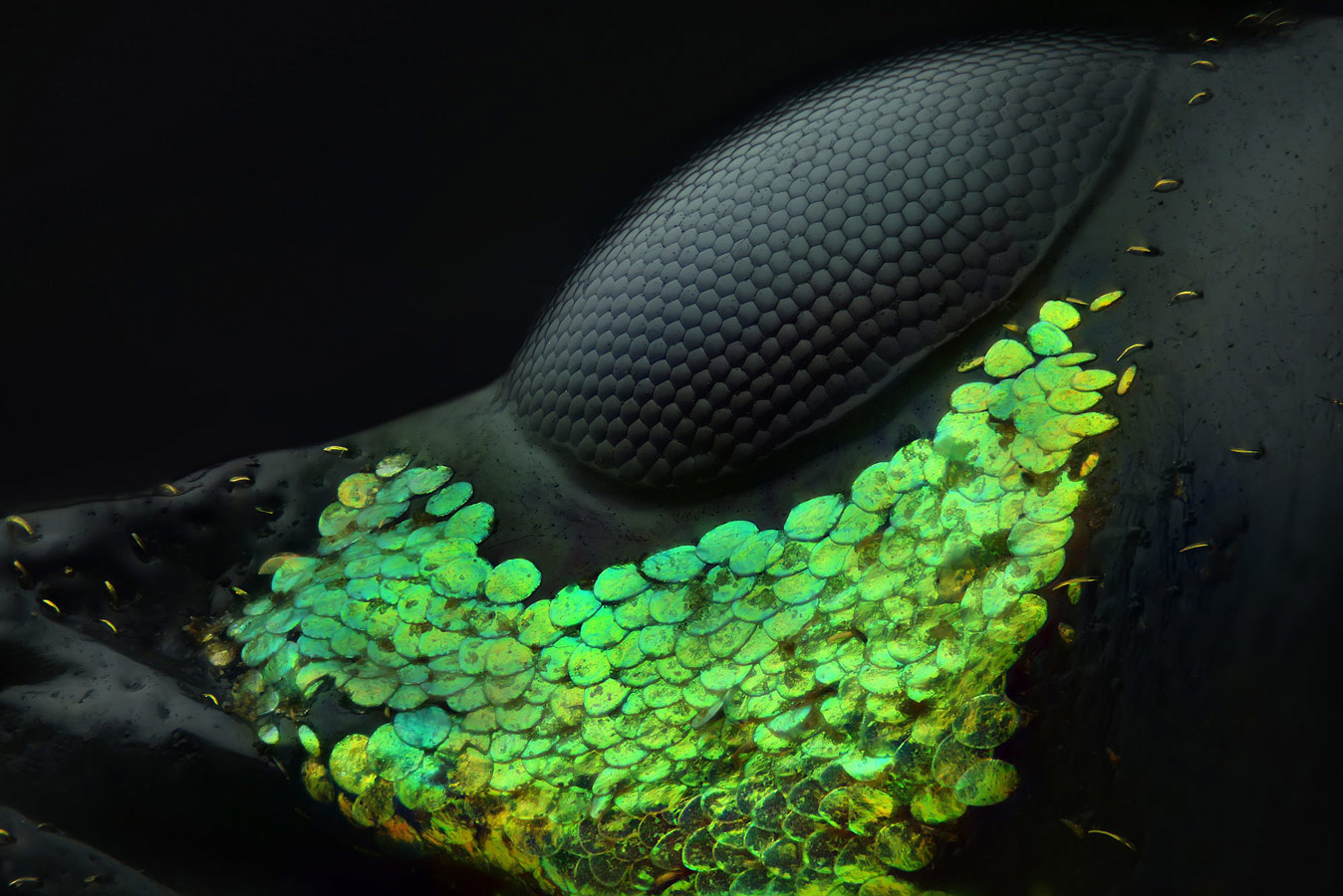
Nikon’s Small World 2018 Winners

Nikon’s Small World


Eye of a Metapocyrtus subquadrulifer beetle, © Yousef Al Habshi, Abu Dhabi, United Arab Emirates, 1st Place, Nikon’s Small World - Photomicrography Competition
Eye of a Metapocyrtus subquadrulifer beetle
© Yousef Al Habshi, Abu Dhabi, United Arab Emirates
1st Place
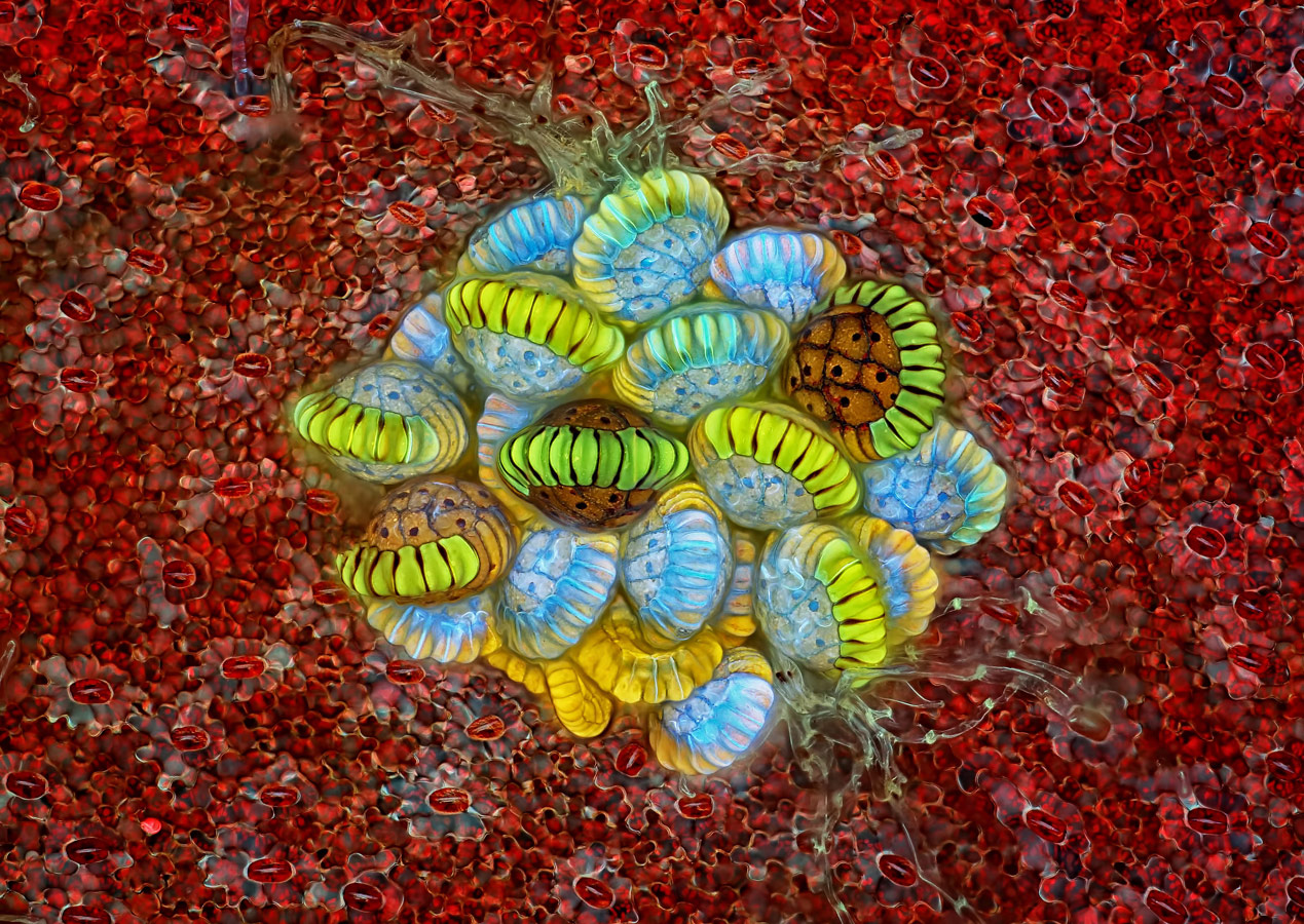
 

Fern sorus (structures producing and containing spores), © Rogelio Moreno Gill, Panama, 2nd Place, Nikon’s Small World - Photomicrography Competition
Fern sorus (structures producing and containing spores)
© Rogelio Moreno Gill, Panama
2nd Place
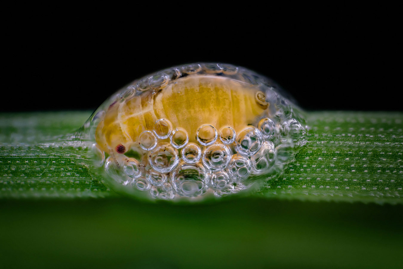
 

Spittlebug nymph in its bubble house, © Saulius Gugis, Naperville, Illinois, USA, 3rd Place, Nikon’s Small World - Photomicrography Competition
Spittlebug nymph in its bubble house
© Saulius Gugis, Naperville, Illinois, USA
3rd Place


Peacock feather section, © Can Tunçer, İzmir, Turkey, 4th Place, Nikon’s Small World - Photomicrography Competition
Peacock feather section
© Can Tunçer, İzmir, Turkey
4th Place
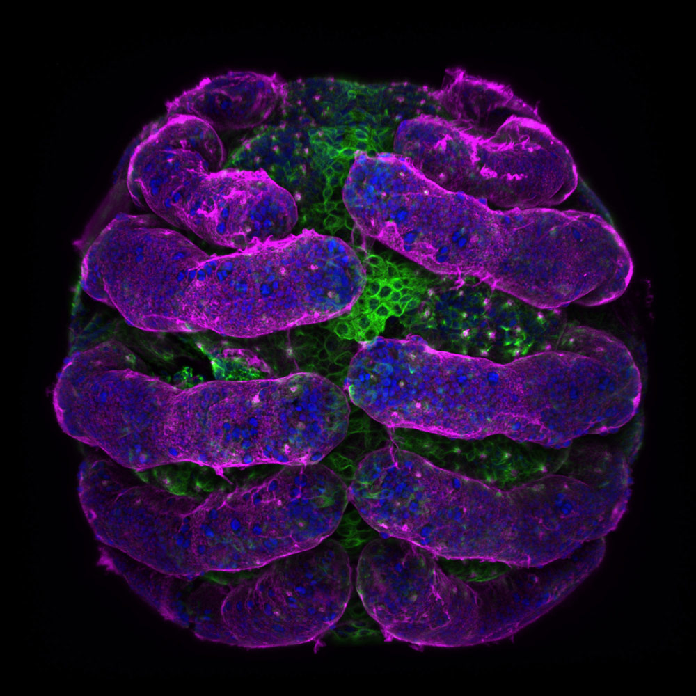
 

Parasteatoda tepidariorum (spider embryo) stained for embryo surface (pink), nuclei (blue) and microtubules (green), © Dr. Tessa Montague, Harvard University, Department of Molecular and Cellular Biology, Cambridge, Massachusetts, USA, 5th Place, Nikon’s Small World - Photomicrography Competition
Parasteatoda tepidariorum (spider embryo) stained for embryo surface (pink), nuclei (blue) and microtubules (green)
© Dr. Tessa Montague, Harvard University, Department of Molecular and Cellular Biology, Cambridge, Massachusetts, USA
5th Place
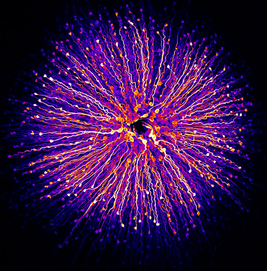
 

Primate foveola (central region of the retina), © Hanen Khabou, Vision Institute, Department of Therapeutics, Paris, France, 6th Place, Nikon’s Small World - Photomicrography Competition
Primate foveola (central region of the retina)
© Hanen Khabou, Vision Institute, Department of Therapeutics, Paris, France
6th Place
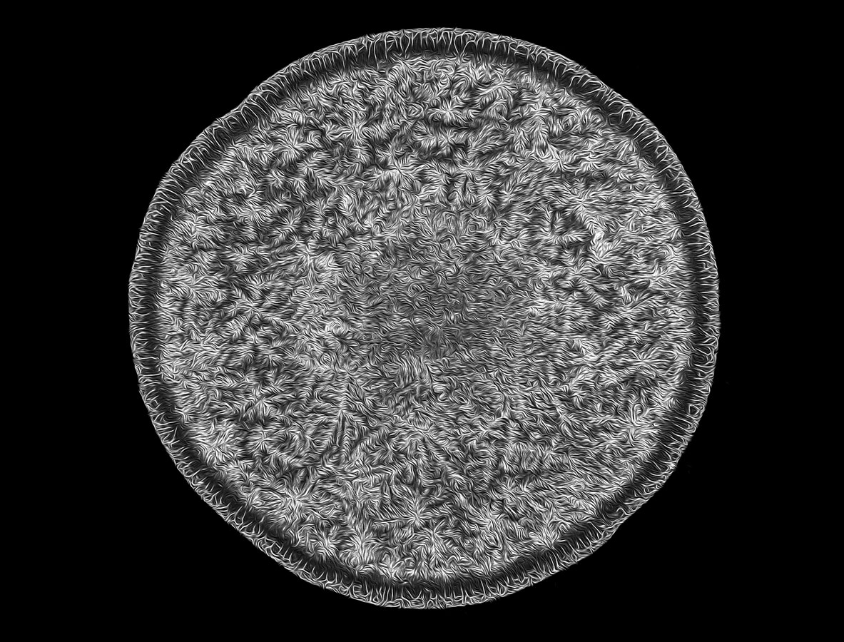
 

Human tear drop, © Norm Barker, Johns Hopkins School of Medicine, Department of Pathology & Art as Applied to Medicine, Baltimore, Maryland, USA, 7th Place, Nikon’s Small World - Photomicrography Competition
Human tear drop
© Norm Barker, Johns Hopkins School of Medicine, Department of Pathology & Art as Applied to Medicine, Baltimore, Maryland, USA
7th Place
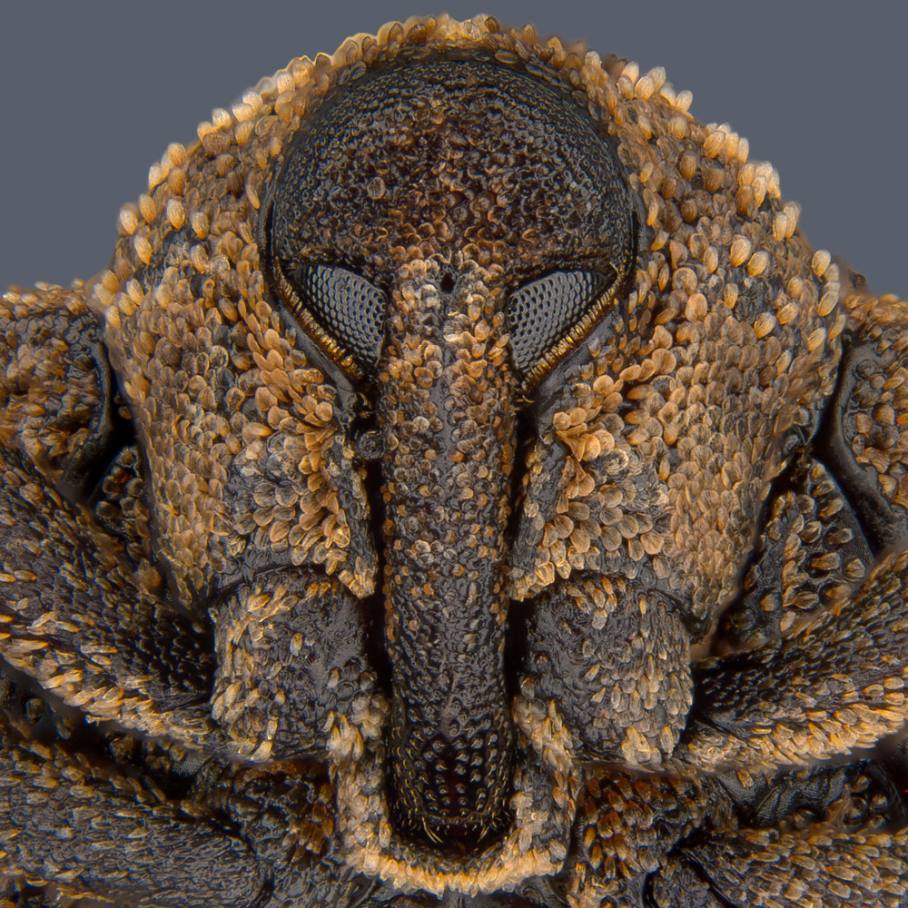
 

Portrait of Sternochetus mangiferae (mango seed weevil), © Pia Scanlon, Government of Western Australia, Department of Primary Industries and Regional Development, South Perth, Western Australia, Australia, 8th Place, Nikon’s Small World - Photomicrography Competition
Portrait of Sternochetus mangiferae (mango seed weevil)
© Pia Scanlon, Government of Western Australia, Department of Primary Industries and Regional Development, South Perth, Western Australia, Australia
8th Place
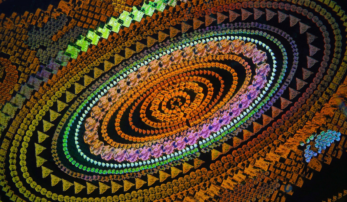
 

Security hologram, © Dr. Haris Antonopoulos, Athens, Greece, 9th Place, Nikon’s Small World - Photomicrography Competition
Security hologram
© Dr. Haris Antonopoulos, Athens, Greece
9th Place
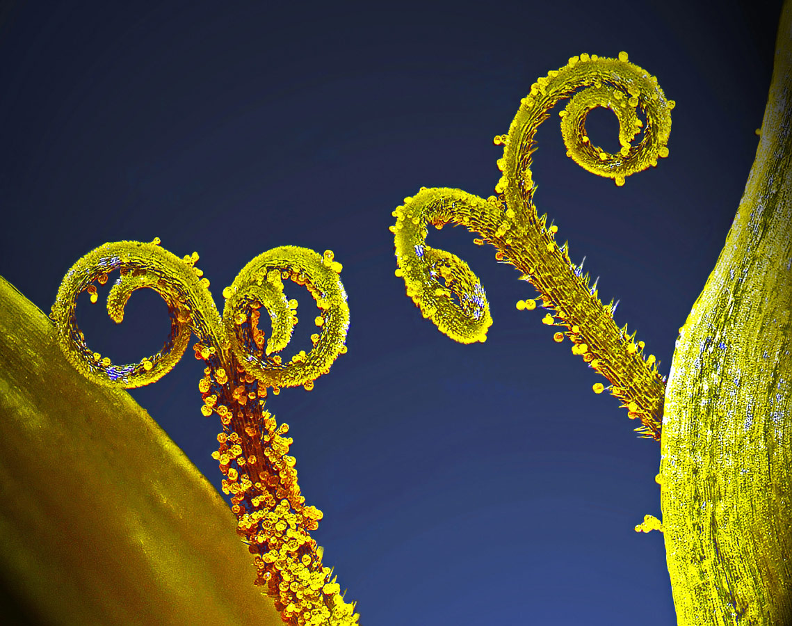
 

Stalks with pollen grains, © Dr. Csaba Pintér, University of Pannonia, Georgikon Faculty, Department of Plant Protection, Keszthely, Hungary, 10th Place, Nikon’s Small World - Photomicrography Competition
Stalks with pollen grains
© Dr. Csaba Pintér, University of Pannonia, Georgikon Faculty, Department of Plant Protection, Keszthely, Hungary
10th Place
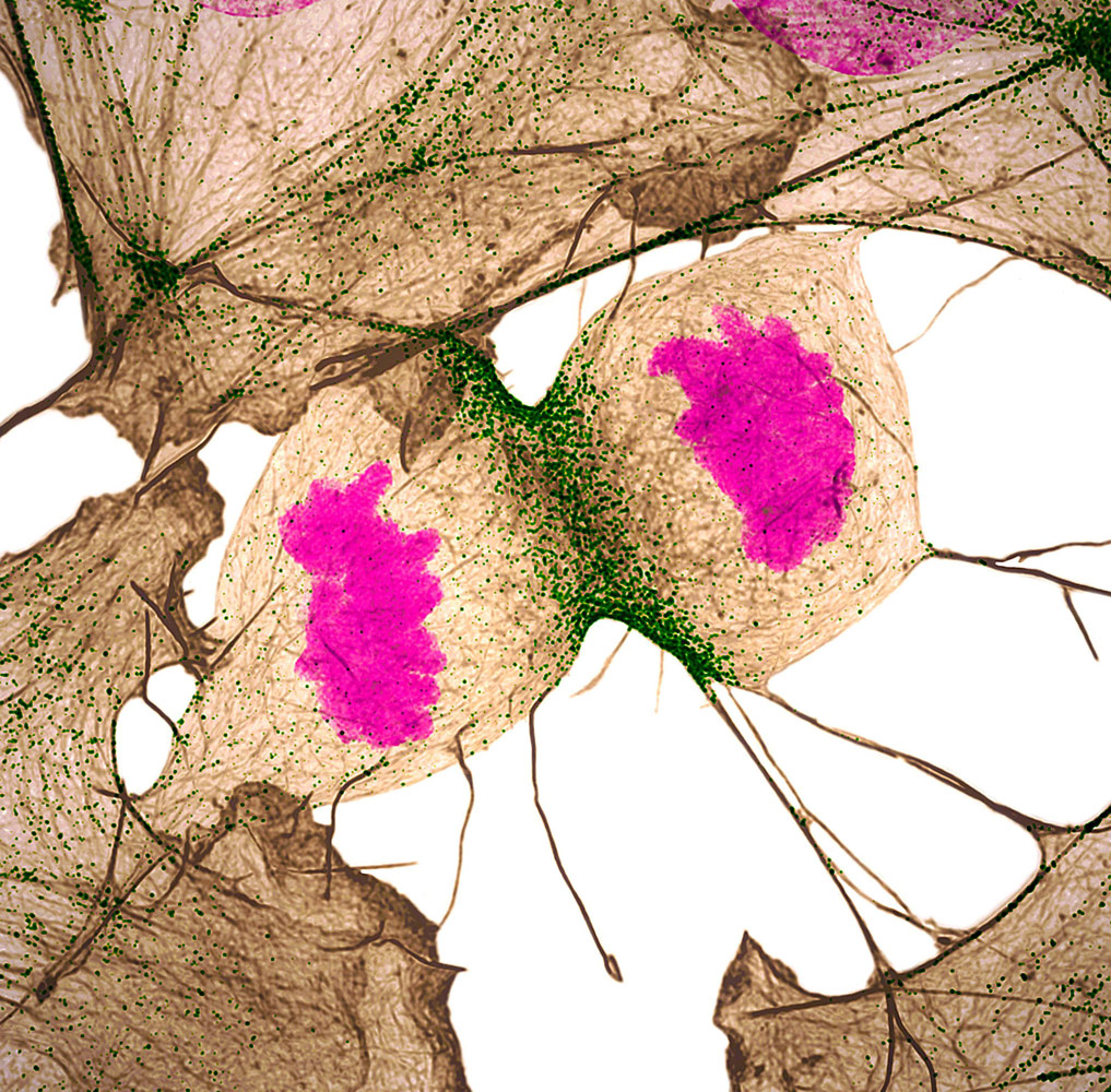
 

Human fibroblast undergoing cell division, showing actin (gray), myosin II (green) and DNA (magenta), © Nilay Taneja, Dr. Dylan Burnette, Vanderbilt University, Department of Cell and Developmental Biology, Nashville, Tennessee, USA, 11th Place, Nikon’s Small World - Photomicrography Competition
Human fibroblast undergoing cell division, showing actin (gray), myosin II (green) and DNA (magenta)
© Nilay Taneja, © Dr. Dylan Burnette, Vanderbilt University, Department of Cell and Developmental Biology, Nashville, Tennessee, USA
11th Place
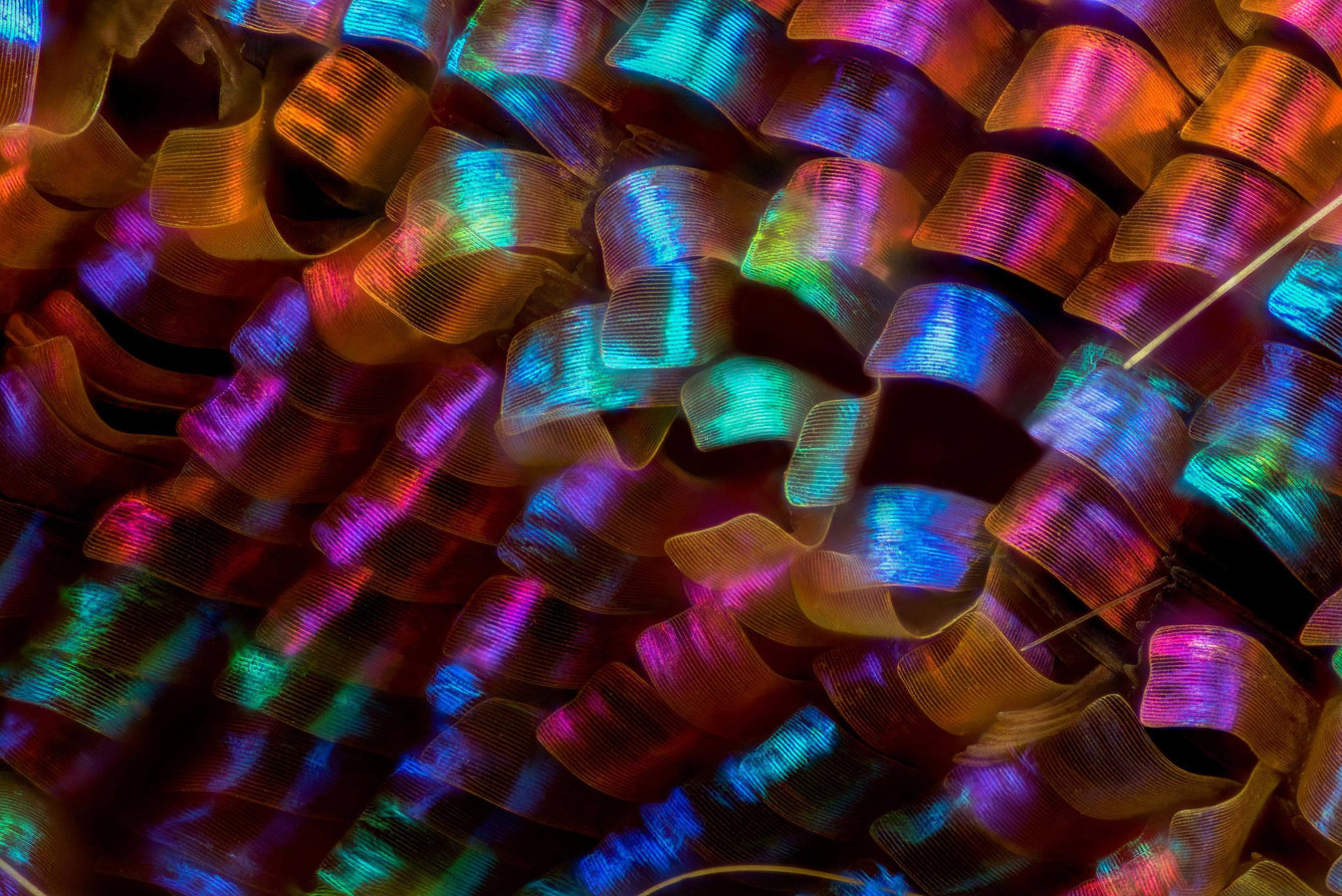
 

Urania ripheus (butterfly) wing scales, © Luciano Andres Richino, Punto NEF Photography, Ramos Mejia, Buenos Aires Province, Argentina, 12th Place, Nikon’s Small World - Photomicrography Competition
Urania ripheus (butterfly) wing scales
© Luciano Andres Richino, Punto NEF Photography, Ramos Mejia, Buenos Aires Province, Argentina
12th Place
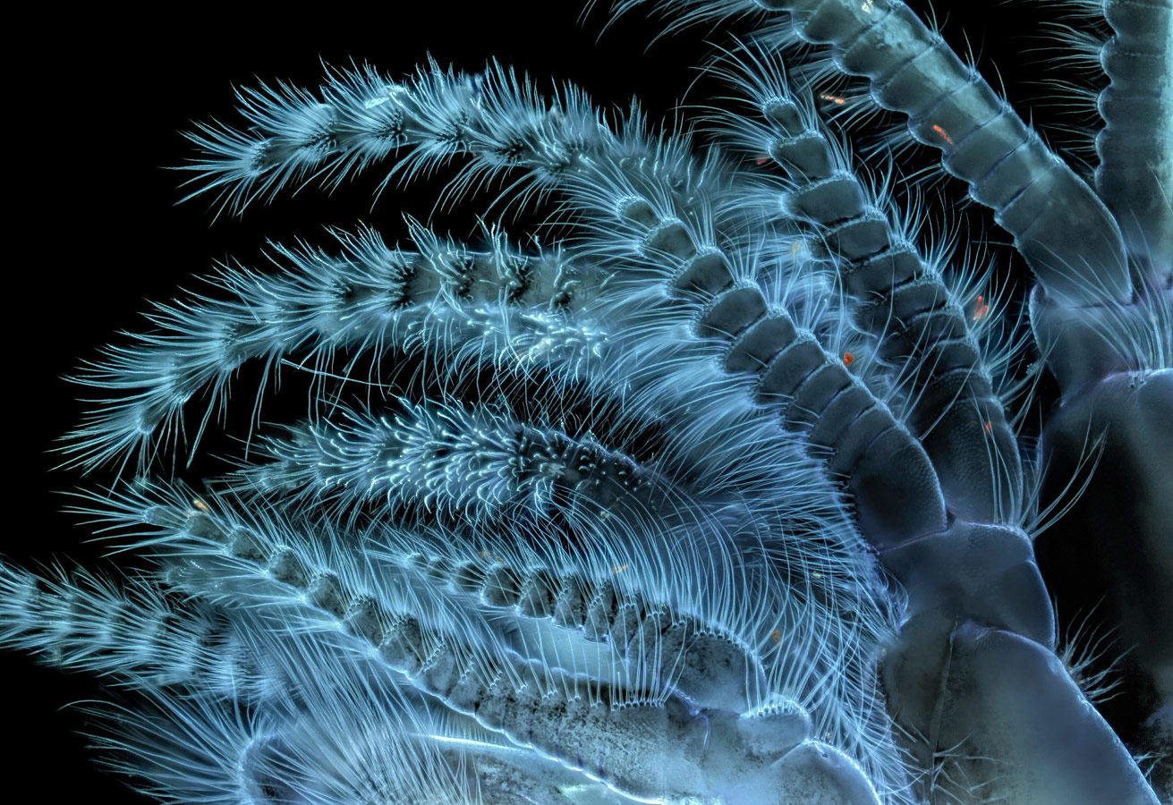
 

Balanus glandula (acorn barnacle), © Charles B. Krebs, Charles Krebs Photography, Issaquah, Washington, USA, 13th Place, Nikon’s Small World - Photomicrography Competition
Balanus glandula (acorn barnacle)
© Charles B. Krebs, Charles Krebs Photography, Issaquah, Washington, USA
13th Place
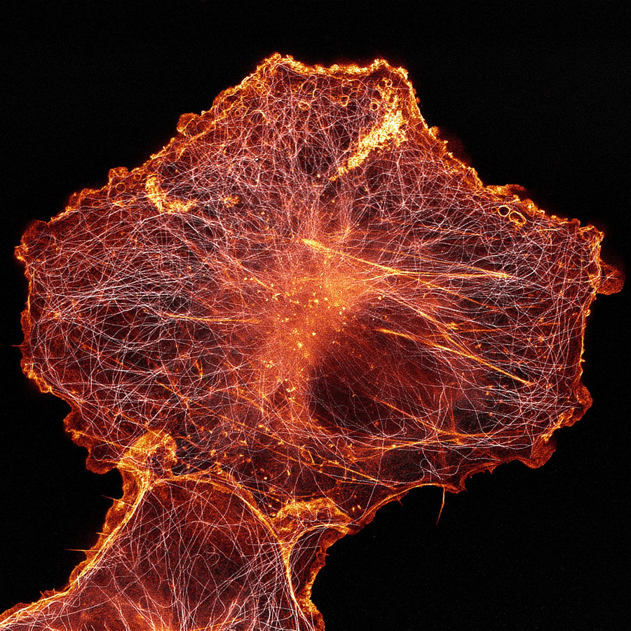
 

African green monkey cell (COS-7) stained for actin and microtubules, © Andrew Moore, © Dr. Erika Holzbaur, University of Pennsylvania, Department of Physiology, Philadelphia, Pennsylvania, USA, 14th Place, Nikon’s Small World - Photomicrography Competition
African green monkey cell (COS-7) stained for actin and microtubules
© Andrew Moore, © Dr. Erika Holzbaur, University of Pennsylvania, Department of Physiology, Philadelphia, Pennsylvania, USA
14th Place
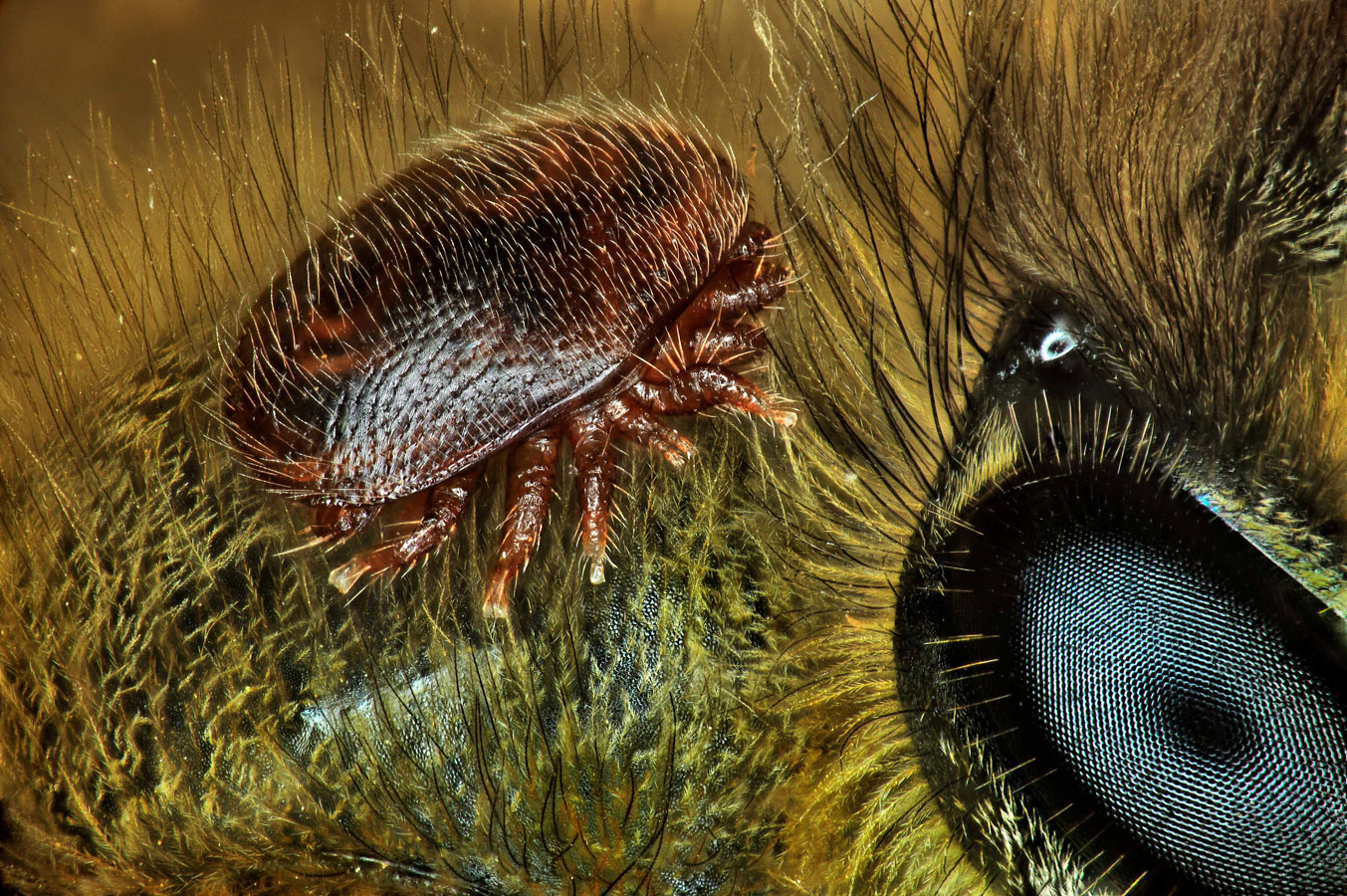
 

Varroa destructor (mite) on the back of Apis mellifera (honeybee), © Antoine Franck, CIRAD - Agricultural Research for Development, Saint Pierre, Réunion Island, France, 15th Place, Nikon’s Small World - Photomicrography Competition
Varroa destructor (mite) on the back of Apis mellifera (honeybee)
© Antoine Franck, CIRAD – Agricultural Research for Development, Saint Pierre, Réunion Island, France
15th Place
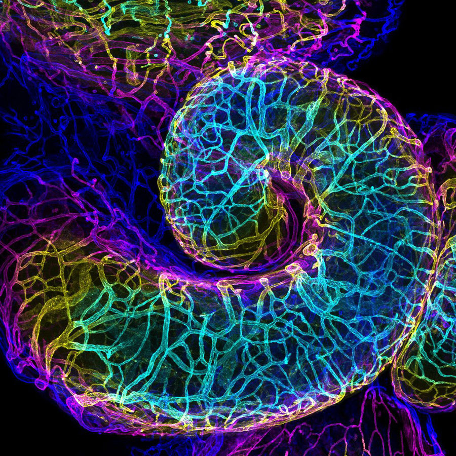
 

Mouse oviduct vasculature, © Dr. Amanda D. Phillips Yzaguirre, Salk Institute for Biological Studies, La Jolla, California, USA, 16th Place, Nikon’s Small World - Photomicrography Competition
Mouse oviduct vasculature
© Dr. Amanda D. Phillips Yzaguirre, Salk Institute for Biological Studies, La Jolla, California, USA
16th Place
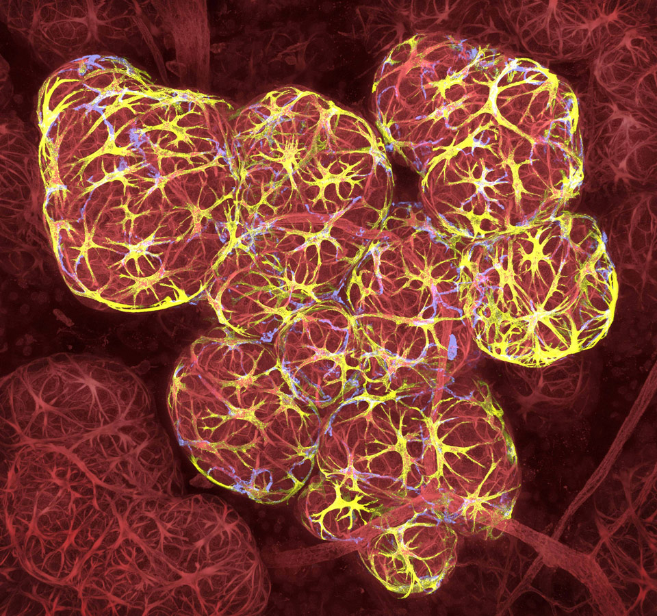
 

Breast tissue in lactation: Milk filled spheres (red) surrounded by tiny muscle cells that squeeze out milk (yellow) and immune cells that monitor for infection (blue), © Caleb Dawson, The Walter and Eliza Hall Institute of Medical Research, Department of Stem Cells and Cancer, Melbourne, Australia, 17th Place, Nikon’s Small World - Photomicrography Competition
Breast tissue in lactation: Milk filled spheres (red) surrounded by tiny muscle cells that squeeze out milk (yellow) and immune cells that monitor for infection (blue)
© Caleb Dawson, The Walter and Eliza Hall Institute of Medical Research, Department of Stem Cells and Cancer, Melbourne, Australia
17th Place
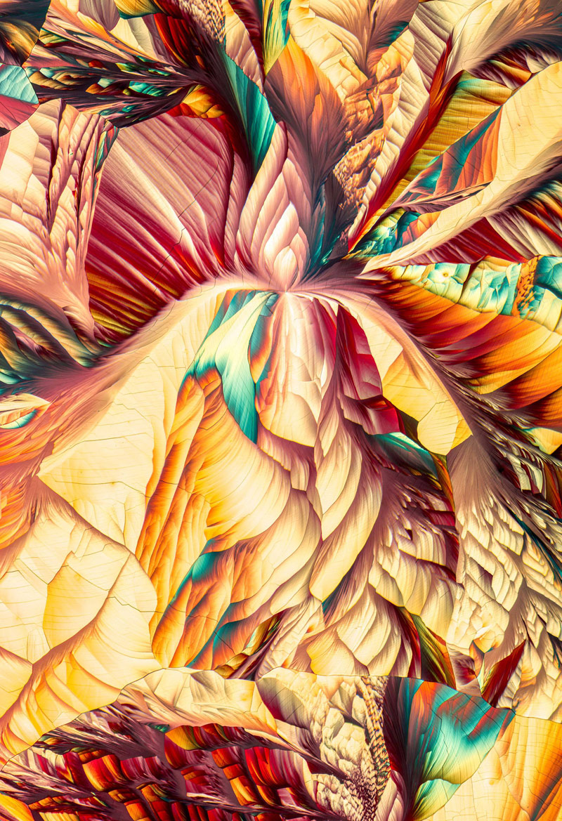
 

Amino acid crystals (L-glutamine and beta-alanine), © Justin Zoll, Justin Zoll Photography, Ithaca, New York, USA, 18th Place, Nikon’s Small World - Photomicrography Competition
Amino acid crystals (L-glutamine and beta-alanine)
© Justin Zoll, Justin Zoll Photography, Ithaca, New York, USA
18th Place
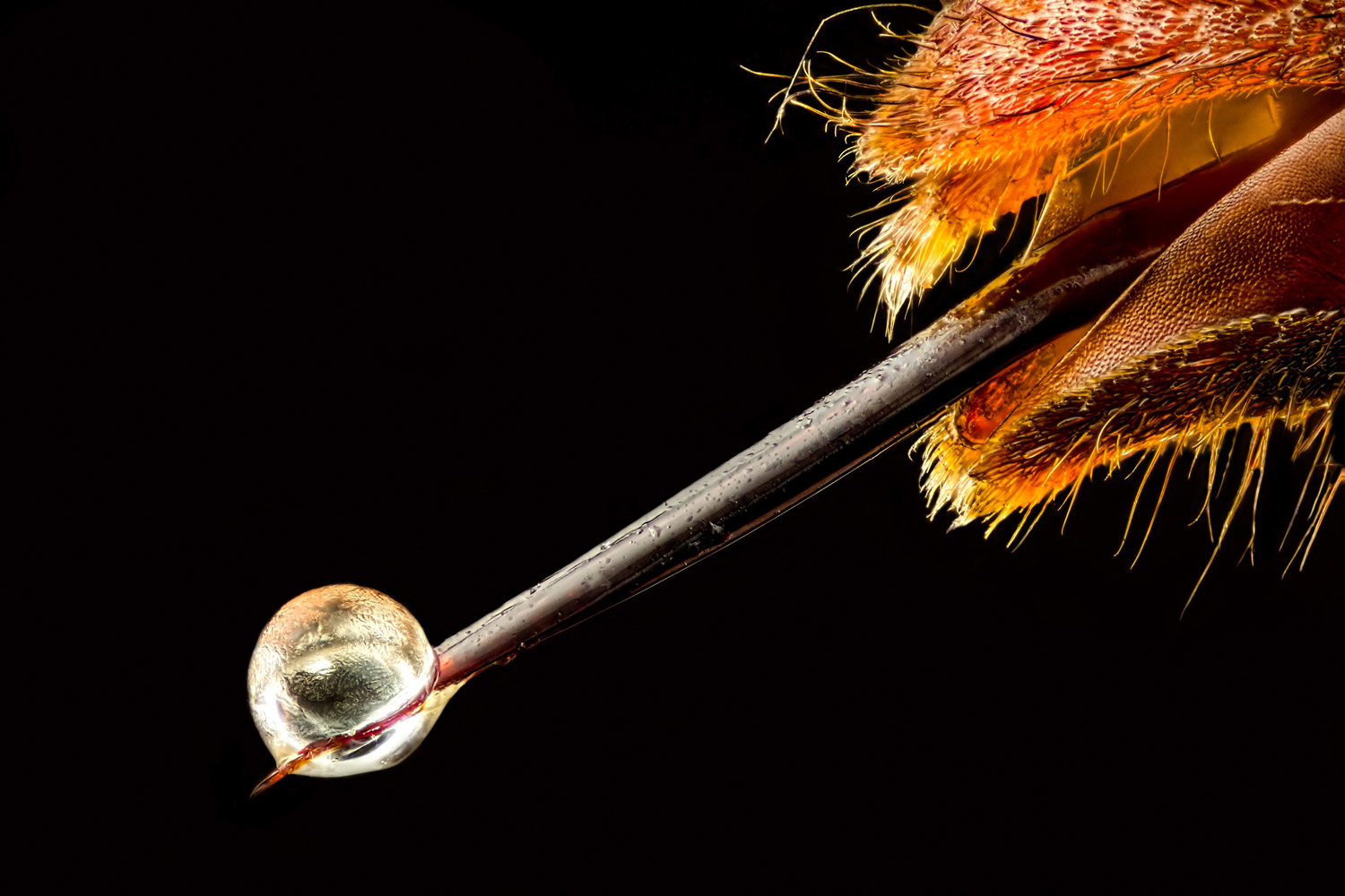
 

Vespa velutina (Asian hornet) with venom on its stinger, © Pierre Anquet, La Tour-du-Crieu, Ariège, France, 19th Place, Nikon’s Small World - Photomicrography Competition
Vespa velutina (Asian hornet) with venom on its stinger
© Pierre Anquet, La Tour-du-Crieu, Ariège, France
19th Place


Human retina, © Dr. Nicolás Cuenca, © Isabel Ortuño-Lizarán, University of Alicante, Department of Physiology, Genetics and Microbiology, San Vicente del Raspeig, Alicante, Spain, 20th Place, Nikon’s Small World - Photomicrography Competition
Human retina
© Dr. Nicolás Cuenca, © Isabel Ortuño-Lizarán, University of Alicante, Department of Physiology, Genetics and Microbiology, San Vicente del Raspeig, Alicante, Spain
20th Place


Nikon’s Small World


Share:

Rating
Share to friends
Pixcontests – Photo Contests of the World